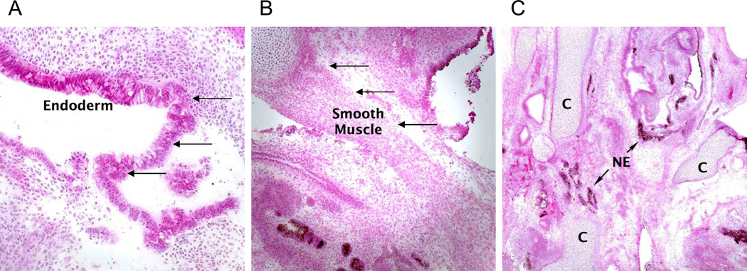
Figure 4. Non-hematopoietic differentiation of RDEB iPS cells.
Histological examination of mature teratoma from an immunodeficient mice injected with KC RDEB iPS cells revealed (A) columnar epithelium of endodermal origin (arrows), (B) smooth muscle of mesodermal origin (arrows), and (C) melanocytes of ectodermal origin (arrows). NE, neuroectoderm; C, cartilage. Similar mature teratomas with contribution of ectodermal-, mesodermal-, and endodermal-derived cells formed after injection of KC WT iPS cells (data not shown). Magnification 20×. Hematoxylin-eosin stain.

