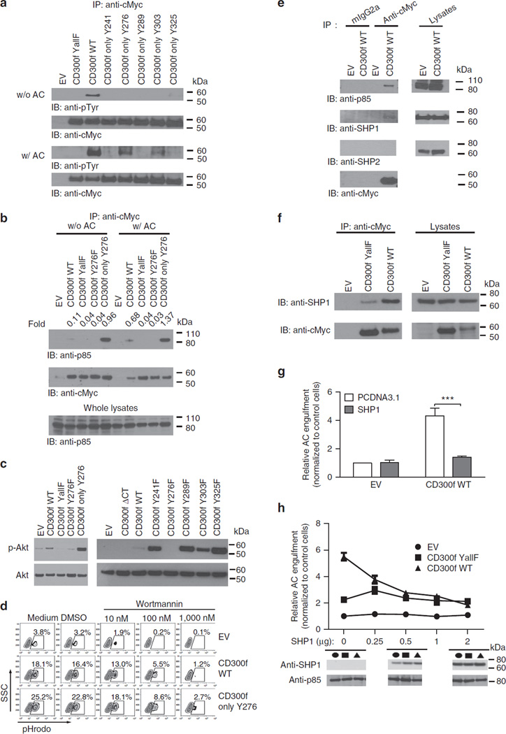
Figure 4. CD300f associates with p85α subunit of PI3K and SHP1 to regulate the phagocytosis of AC.
(a–c) L929 cells transduced with EV or cMyc-tagged CD300f constructs were incubated with (a–c) or without (a,b) AC at 4°C for 10 min, then transferred to 37 °C for 15 min before lysis. Immunoprecipitation was performed with anti-cMyc Ab (a,b), and blotting with antibodies against phosphotyrosine (a), p85 (b) or cMyc (a,b) (loading control). The presence of PI3K subunit was verified by anti-p85 immunoblotting of whole lysates (b); anti-phospho-Akt or anti-Akt (loading control) blots were performed on whole lysates (c). (d) L929 cells transduced with the indicated constructs were preincubated with medium or DMSO (controls), or Wortmannin for 30 min at 37 °C, followed by 2 h incubation with pHrodo-labelled AC. The percentage of cells containing AC was determined as in Fig. 1. (e) J774 cells transduced with EV or cMyc-tagged CD300f WT were treated with pervanadate for 15 min at 37 °C, lysed, immunoprecipitated with anti-cMyc or isotype control antibodies and then blotted with anti-p85, anti-SHP1, anti-SHP2 or anti-cMyc antibodies. Lysates blotted with anti-p85, anti-SHP1 or anti-SHP2 antibodies served as controls. (f) The indicated cMyc-tagged CD300f constructs were co-transfected with SHP1 into HeLa cells. Cells, treated with pervanadate, were lysed, immunoprecipitated with anti-cMyc Ab and blotted with anti-SHP1 or anti-cMyc antibodies. Cell lysates were blotted with anti-SHP1 or anti-cMyc for SHP1 or CD300f presence. (g) EV or CD300f WT was co-transfected with pCDNA3.1 or SHP1 into HeLa cells. Cells were incubated with pHrodo-labelled AC for 2 h, and phagocytosis efficiency was analysed as in Fig. 1, and expressed as relative values normalized to EV- and pCDNA3.1-transfected control cells. The graph shows means with s.e.m. (error bars) from three experiments. Asterisks indicate statistical significance (***P<0.001, Student’s t-test). (h) The indicated amounts of SHP1-encoding vector were co-transfected with EV or CD300f constructs into HeLa cells. The ability of cells to phagocytize pHrodo-labelled AC was analysed as in (g) and plotted versus SHP1 transfection level. The graph shows means with s.e.m. (error bars) from three experiments. The images below the graph show representative anti-SHP1 blots of lysates from cells transfected with 0, 0.5 or 2 µg of SHP1-encoding vector. Anti-p85 immunoblotting served as loading control.

