
Figure 2.
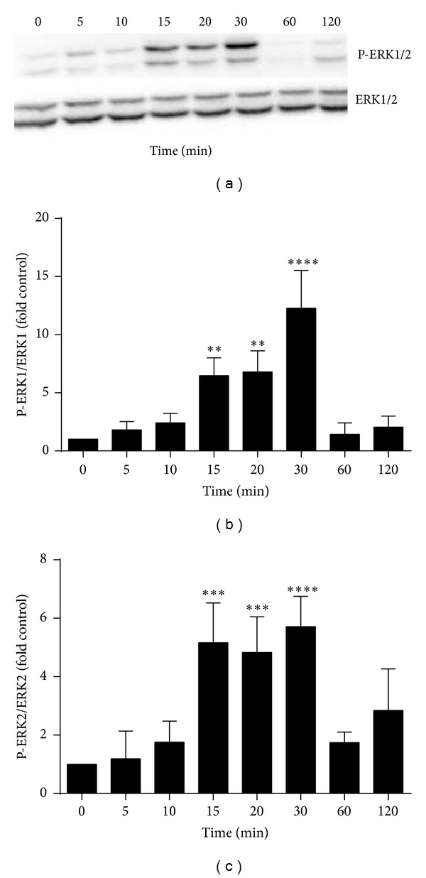
LPS induces phosphorylation of ERK1/2 in a time-dependent manner. RAW 264.7 cells were incubated with 100 ng/mL LPS for 0–24 h. Total cell lysates were examined by western blot with anti-ERK1/2 and phosphor-ERK1/2 (P-ERK) antibodies. (a) Results shown are representative blots from three different experiments. (b and c) The histograms represent the optical density of phosphor-ERK1/2: total ERK1/2 expressed as control fold increase. Results shown are the mean ± SD of the three experiments. ∗∗∗∗P < 0.0001, ∗∗∗P < 0.001, and ∗∗P < 0.01 compared to control.