
Figure 1.
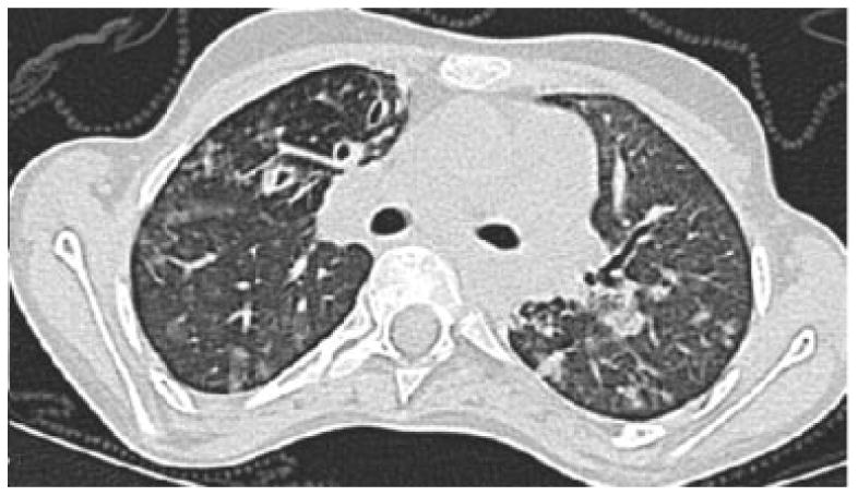
CT image of 17 year old female with A-T and history of recurrent pneumonias, dysphagia and bronchiectasis. This single CT image demonstrates right middle lobe bronchiectasis and bronchial wall thickening, peripheral tree in bud opacities and scattered areas of ground glass opacity.