
Figure 3.
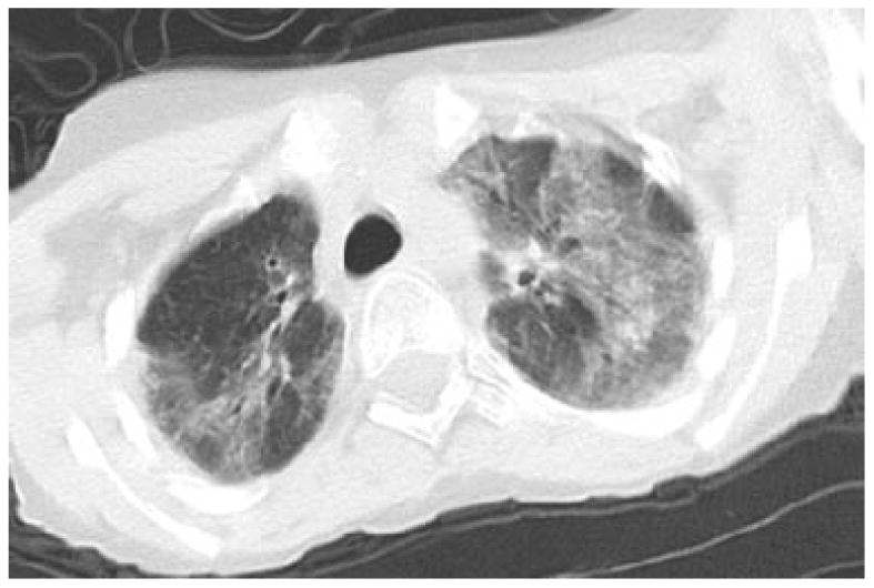
CT image of 14 year old boy with respiratory failure following chemotherapy for lymphoma. This CT image, through the upper lungs demonstrates mosaic attenuation with bilateral geographic areas of predominantly ground glass opacity. Several dilated bronchi are seen in the right upper lobe. Note that this image shows some blurring from motion, to decrease radiation exposure, images that are diagnostic but not ideal should not be repeated in people with A-T.