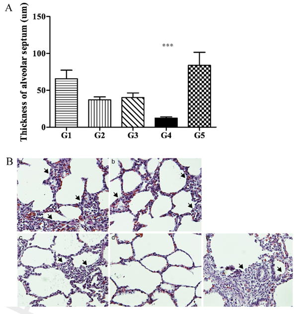
Fig. 3. Histopathological examination.

The graph depicts the thicknesses of the alveolar septa (A), and the photomicrographs (B) show hematoxylin and eosin-stained sections of the cranial lobes of the lungs following the in vivo experiments. The piglets were injected once with wt virus (G1, a), twice with wt virus (G2, b), once with mutant virus (G3, c), twice with mutant virus (G4, d), or sterile media (G5, e). Asterisks indicate significant differences (P < 0.05) between the indicated pairs of groups. The arrows indicate the localized regions of severe interstitial pneumonia and thickened alveolar septa. Magnification: ×200.
