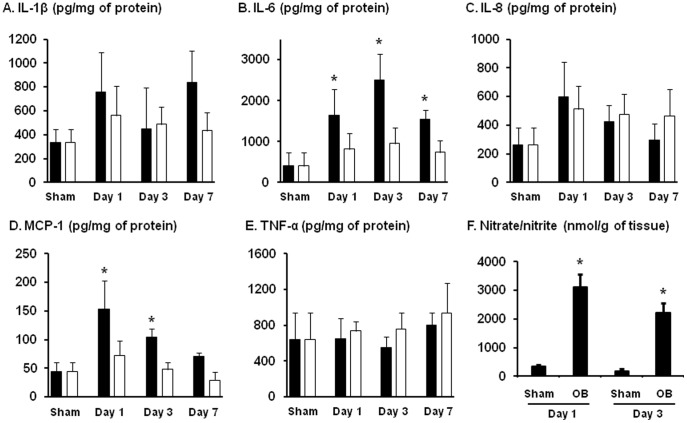
Figure 3. Detection of IL-1β (A), IL-6 (B), IL-8 (C), MCP-1 (D), TNF-α (E) and nitric oxide metabolites (F) in the colonic muscularis externae in rat model of partial colon obstruction.
Rats were euthanized on day 1, 3, and 7 after the operation. Muscularis externae was isolated from the colonic segment oral (black bar) and aboral (white bar) to obstruction band for protein extraction and EIA quantification of cytokines and chemokines (A to E). Y-axis unit for each mediator from A to E is pg/mg of protein. The NO metabolites (nitrate/nitrite) from the colonic muscularis externae were detected in the incubation medium as described in the Methods (F). The Y-axis unit for F is nmol/g of tissue. N = 5 or 6 rats in each group. * p<0.05 compared to sham control.

