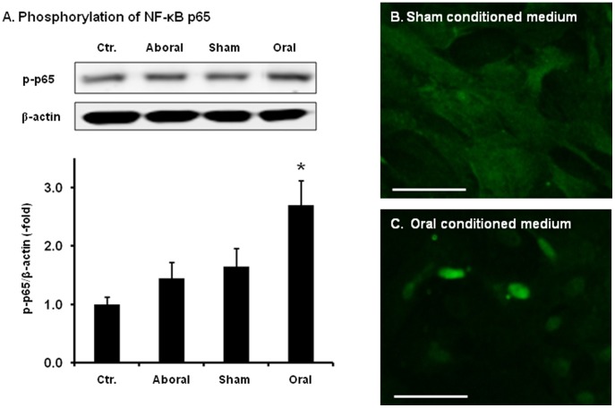
Figure 4. The phosphorylation and translocation of NF-κB p65 in RCCSMCS cultured with conditioned medium.
(A) The NF-κB p65 phosphorylation was detected by Western blot with specific phosphor-p65 antibody. Cells were treated with conditioned media (1∶2 dilution) for 30 min. The p65 phosphorylation level increased 2.7±0.5 folds when naïve RCCSMCs were treated for 30 minutes with conditioned medium from the incubation of the oral segment (N = 4, * p<0.05 vs. blank control). (B) Immunofluorescence staining of p65 in RCCSMCs treated with conditioned medium from the sham tissue for 30 min. (C). Immunofluorescence staining of p65 in RCCSMCs treated with conditioned medium from the oral tissue for 30 min (Images shown are representative of 3 repetitions. Bars = 100 µm.)

