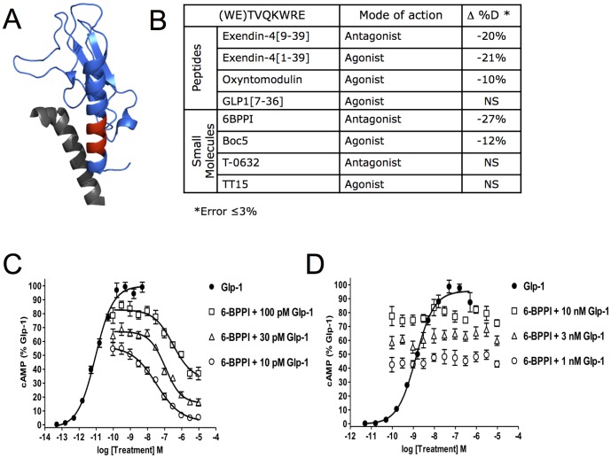
Figure 4. Changes to HDX of nGLP-1R in the Presence of Peptide and Small Molecule Ligands.
A) The WETVQKWRE peptide of nGLP-1R where protection was observed is shown in red on the crystal structure pdb:3C5T. Deuterium exchange was measured in the presence of DDM/CHS micelles as indicated in the Experimental section The exendin-4[9-39] peptide ligand is colored gray. B) Table showing the average change in % Deuterium in this same nGLP-1R peptide (WETVQKWRE) for four peptide ligands and four proposed small molecule ligands. GLP-1 induced cAMP accumulation was quantified in HEK293 cells transfected with plasmids expressing either (C) the GLP-1R or (D) a GIPR/GLP-1R chimeric protein where the GLP-1R 7TM domains are fused in-frame with the GIP-R ectodomain. Dose response curves of GLP-1 were generated and robust cAMP accumulation was observed for the GLP-1R (EC50 = 9.0 pM) and GIPR/GLP-1R (1.3 nM). The ability of 6-BPPI to antagonize GLP-1 signaling was measured by treatment of cells with a dose-response of 6-BPPI in the presence of EC50 to EC80 concentrations of GLP-1. Dose-dependent blockade of GLP-1R signalling by 6-BPPI (100 pM GLP-1, IC50 = 310 nM; 30 pM GLP-1, IC50 = 110 nM, 10 pM GLP-1, IC50 = 33 nM) but not GIPR/GLP-1R was observed.

