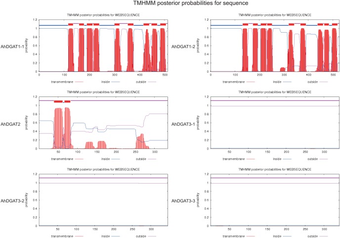
Figure 3. Predicted transmembrane domain for peanut DGAT1, DGAT2 and DGAT3 sequences.
The TMHMM web tools (http://www.cbs.dtu.dk/services/TMHMM-2.0/) plot the probability of the ALDH sequence forming a transmembrane helix (0–1.2 on the y-axis). The regions predicted to form transmembrane helix (or helices) are shown in red, while regions of all sequences predicted to be located inside or outside the membrane are shown in blue and pink, respectively. Nine predicted transmembrane helices were identified for AhDGAT1-1 and AhDGAT1-2 sequences, while two transmembrane helix were observed for AhDGAT2 sequences. No transmembrane helice was identified for AhDGAT3-1, AhDGAT3-2 and AhDGAT3-3 sequences.

