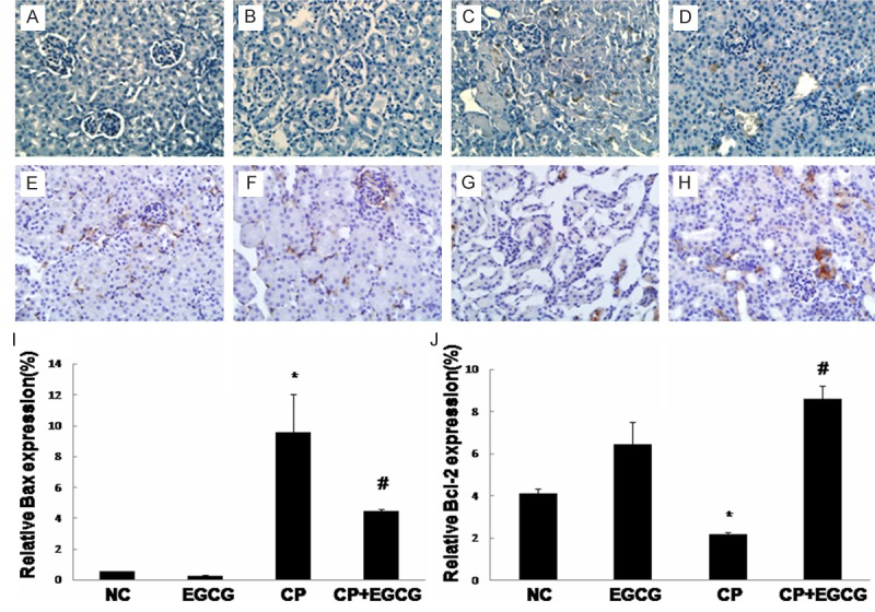
Figure 4.

Immunohistochemical detection of Bax and Bcl-2: Mice were treated with vehicle (A, E), EGCG (B, F), CP (C, G), and CP+EGCG (D, H), separately. The brown granules represent positively stained cells. Original magnification, ×400. Measurement of the intensity of Bax (I) and Bcl-2 (J) immunostaining. Data are presented as means ± SD. *p<0.05 versus control group; #p<0.05 versus CP group.
