
Figure 2.
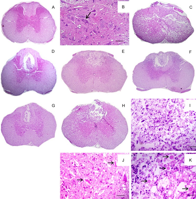
Photograph of rats spinal cord histological sections from an animal subjected to laminectomy (A); detailed view of the gray matter with neuronal integrity (arrow) (B); Photograph of epicenter histological section from an animal subjected to SCI (C); Photograph of cranial histological section from animals subjected to SCI and not treated (D); treated with methylprednisolone (E); methylprednisolone and dantrolene (F); and with dantrolene alone (G); Photograph of caudal histological section from an animal subjected to SCI, with malacia in the dorsal funiculus (H); detailed view of the region of malacia showing gitter cells, cellular debris and inflammatory cells (I); detailed view of the gray matter with degenerated neurons (arrows) (J); and white matter degeneration with axonal swelling and myelin sheaths (arrows) (K) (Figure 3A, C-H: bar=233 mm; Figure 3B, I-K: bar=23 μm.).