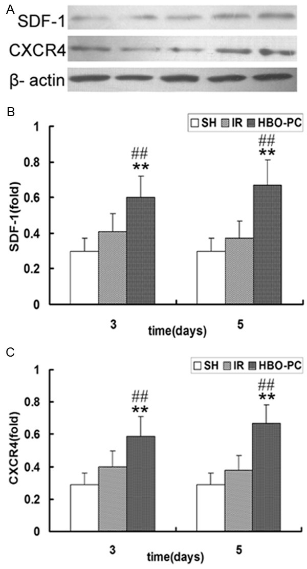
Figure 5.

(A) Immunoblot analysis of SDF-1 and CXCR4 in each group. Western blot analysis of (B) SDF-1 and (C) CXCR4 in the skin flap tissue of each group. SDF-1 and CXCR4 expression in the HBO-PC groups were significantly higher than those in the SH group (**P<0.01). Compared with the same day IR groups, SDF-1 and CXCR4 expression were significantly increased in the HBO-PC groups (##P<0.01). IR, ischemia reperfusion; HBO-PC, hyperbaric oxygen preconditioning; SH, sham-operated.
