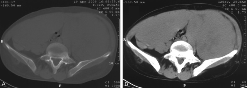
Figure 1.

Abdominal computed tomography. A. Non-enhanced axial CT image demonstrates a large, homogeneous, soft tissue mass about 20 × 15 cm in diameter, occupying the whole abdominal and pelvic cavity and compressing the intraabdominal organs to the caudal side. B. Contrast-enhanced CT shows a tumor with early enhancement in mainly dorsal side of the tumor.
