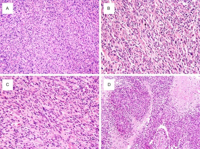
Figure 2.

Spindle cell RMS. (A) The tumor showed high cellularity consisted predominately of spindle cells in long fascicles, arranged in a herringbone pattern, resembling adult-type fibrosarcoma. The spindle cells had elongated and hyperchromatic nuclei, prominent nucleoli and scant cytoplasm, often arranged in a fascicular or herringbone growth pattern. (B) Scattered rhabdomyoblasts containing abundant eosinophilic cytoplasm and eccentrically placed nuclei are admixed with spindled cells. (D) Tumor cells demonstrated a higher mitotic activity and mitotic figures. (E) Spindle cells interspersed with larger geographic areas of necrosis. H & E, original magnification, (A, D), × 200; (B, C), × 400.
