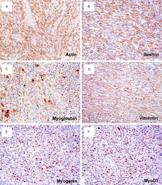
Figure 3.
Immunohistochemistry of spindle cell RMS. Tumor cells are stained positively for muscle specific actin (A), desmin (B), myoglobin (C), and vimentin (D). Scattered tumor cells show a nuclear positivity for myogenin (E) and MyoD1 (F). DAKO Envision peroxidase detection system, (A), × 200; (B-F), × 400.

