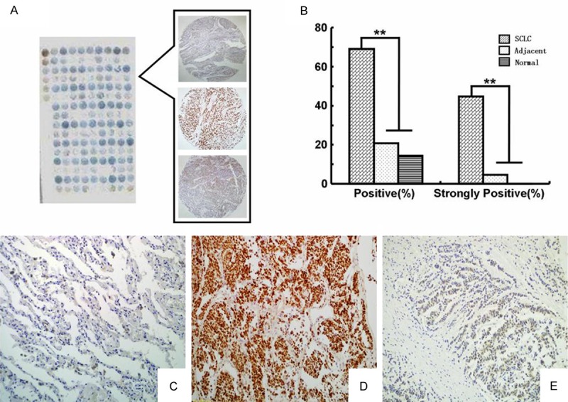
Figure 1.

IHC staining of the DEK protein in SCLC and adjacent non-tumor tissue. DEK protein was detected in tissue microarray (A) by IHC staining. The histogram (B) showed that the positive rate and strongly positive rate of DEK protein expression were significantly higher in SCLC than that in either adjacent non-tumor or normal lung tissues. DEK was negative in normal lung tissue (C), but strongly positive in SCLC with lymph node metastasis (D) and weakly positive in SCLC with non-metastasis (E) (original magnification, (C-E) × 200).
