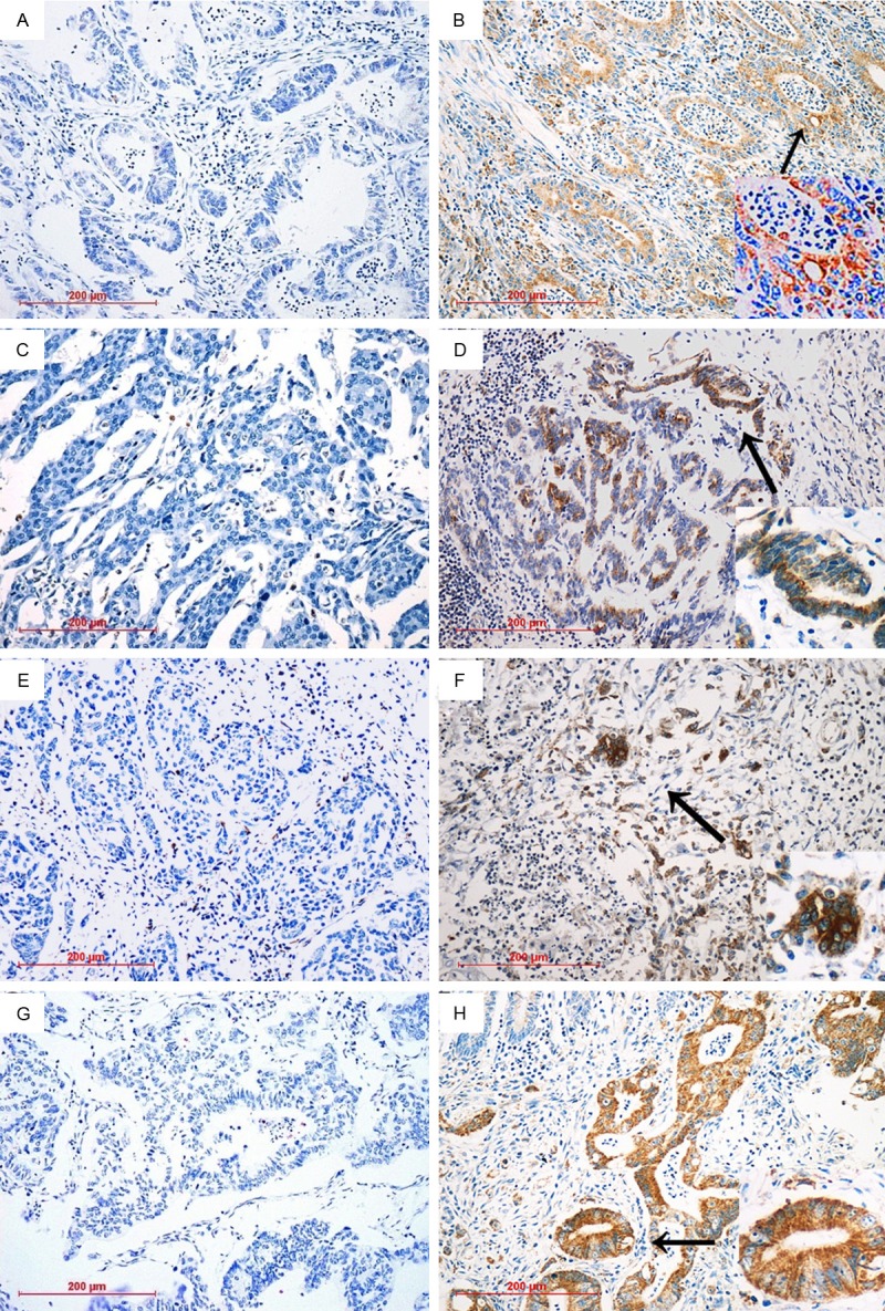
Figure 1.

IHC analysis of cleaved caspase-3 in human cancer smples. Paraffin-embedded tissue sections were stained using an immunoperoxidase method, as described in Materials and methods. Representative images (200×) are shown. A. Representative IHC staining patterns for samples with low cleaved caspase-3 in gastric cancer; B. Representative IHC staining patterns for samples with high levels of cleaved caspase-3 in gastric cancer; C. Representative IHC staining patterns for samples with low cleaved caspase-3 in ovarian cancer; D. Representative IHC staining patterns for samples with high levels of cleaved caspase-3 in ovarian cancer; E. Representative IHC staining patterns for samples with low cleaved caspase-3 in cervical cancer; F. Representative IHC staining patterns for samples with high levels of cleaved caspase-3 in cervical cancer; G. Representative IHC staining patterns for samples with low cleaved caspase-3 in colorectal cancer; H. Representative IHC staining patterns for samples with high levels of cleaved caspase-3 in colorectal cancer. As shown above, intense immunostaining for cleaved caspase-3 protein was mainly presented in the cytoplasm of the tumor cell, and partially localized in the nucleus.
