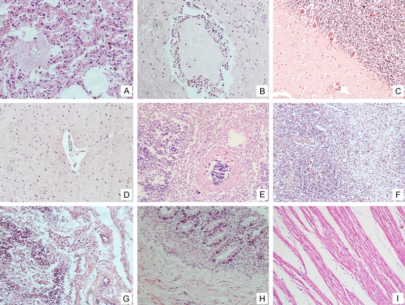
Figure 1.

Histological findings of the main tissues of the autopsy. Lungs manifested as typical severe pulmonary congestion and edema with focal intra-alveolar hemorrhage (A); Sleevelet-like inflammatory cells infiltrated around the blood vessel in the brain. The cerebellum and brain stem showed the inflammatory cell infiltration (B-D); The thymus showed follicular hyperplasia (E); The spleen was congested with phagocytes and mononuclear cells (F); The mesenteric lymph nodes showed mild lymphoid hyperplasia and congestion (G); There was slight histological change in the mucosa of the alimentary tract with only a mild degree of focal monocytes, lymphocytes infiltration (H); The heart showed mild local exudation with a small focus of mononuclear cell infiltration in the myocardium. The myocardial cells were intact without degeneration and rupture (I).
