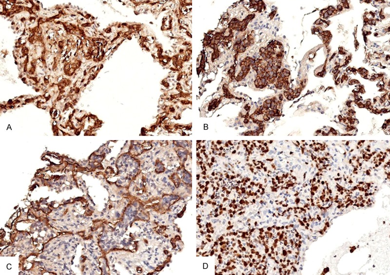
Figure 3.

Immunohistochemical staining of the neoplastic intravascular tumor cells. A: Positive expression of LCA in the intravascular neoplastic cells (400X); B: Positive expression of CD20 in the intravascular neoplastic cells (400X); C: CD34 immunostaining shows the neoplastic lymphocytes located in the alveolar capillaries (400X); D: KI-67 immunostaining highlights the proliferation of intravascular lymphoma cells (400X).
