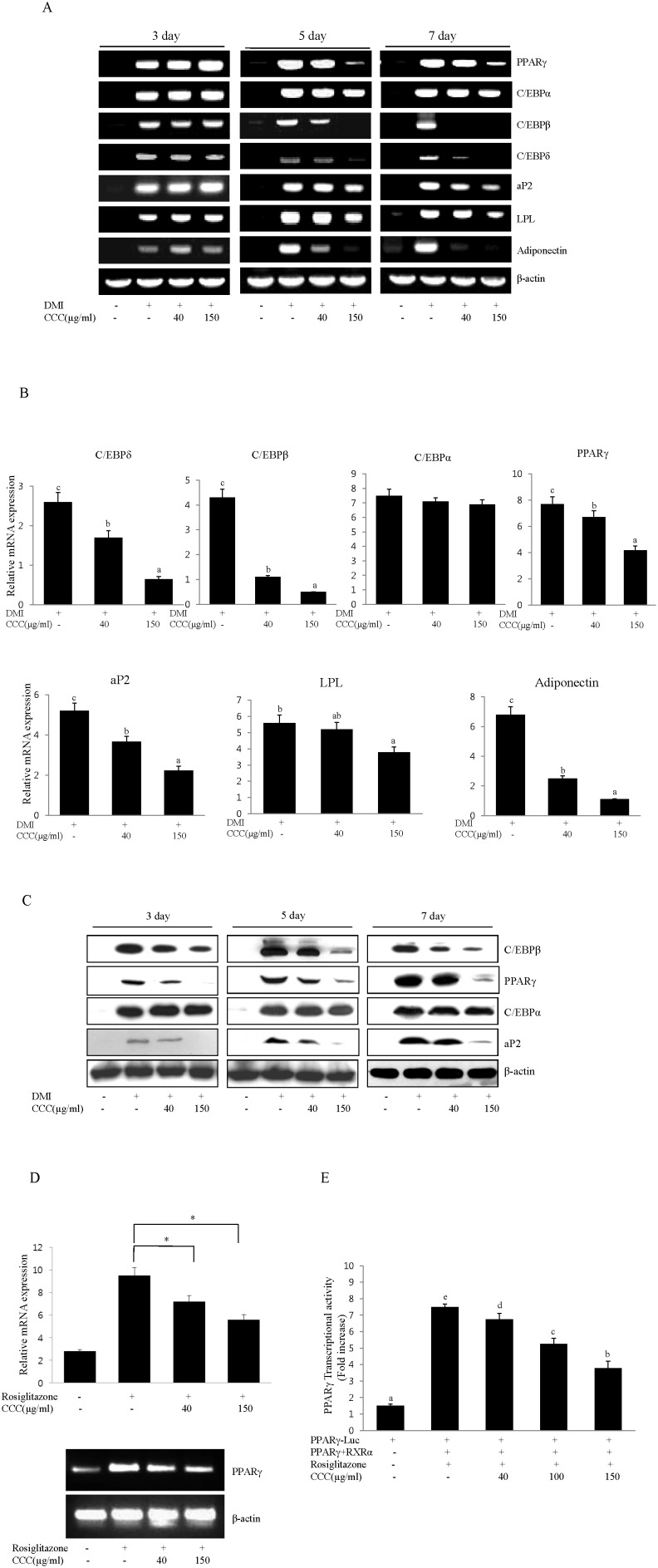
Figure 2. CCC down-regulated the expression of the main adipogenesis and adipocyte-specific transcription factors during adipocyte differentiation in 3T3-L1 preadipocytes.
(A) Effects of CCC on PPARγ, C/EBPα, C/EBPβ, C/EBPδ, aP2, LPL, and adiponectin mRNA expression in 3T3-L1 cells. Confluent 3T3-L1 preadipocytes were induced to differentiate in the presence of different concentrations of CCC (from 0 to 150 µg/ml) for 7 days. Total RNA was isolated from 3T3-L1 adipocytes on days 3, 5 or 7 after the induction of differentiation, and gene expression analysis was performed by RT-PCR. The amplification of β-actin was performed as a loading control. All of the experiments were performed in three independent experiments. (B) The expression levels of adipogenesis and adipocyte-specific genes were determined during adipocyte differentiation at day 7. The mRNA expression of PPARγ, C/EBPα, C/EBPβ, C/EBPδ, aP2, and LPL were normalized using β-actin as a control. The bars with different letters are significantly different (*p<0.05) as determined by Duncan's multiple range test. (C) CCC reduced the protein levels of PPARγ, C/EBPβ, and aP2 genes in 3T3-L1 adipocytes. Total protein was isolated from 3T3-L1 adipocytes on days 3, 5 or 7 after the induction of differentiation. Total cellular proteins were immunoblotted for PPARγ, C/EBPβ, aP2, and β-actin, as indicated. Similar results were obtained from 3 replicates. (D) Effect of CCC on PPARγ mRNA expression in A549 lung cancer cells. A549 lung cancer cells were treated with 40 or 150 µg/ml CCC in the absence or presence of rosiglitazone (10 µM) for 24 h in complete growth medium. Total RNA isolated from A549 cells was subjected to RT-PCR, and all of the gene transcripts were normalized using β-actin as a control. The data represented the mean ± SD of 3 different experiments. *p<0.05, **P<0.01. (E) PPARγ and RXRα were cotransfected with the PPARγ-Luc reporter construct into CHO cells for 24 h. The cells were treated with 100 µM rosiglitazone in the absence or presence of CCC. After 24 h, luciferase activity was assayed. The data are the mean ± SD values of at least three independent experiments. The bars with different letters are significantly different (*p<0.05) as determined by Duncan's multiple range test.

