
Fig. 3.
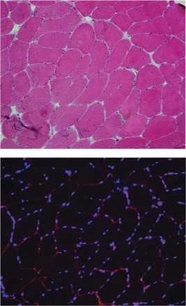
Top: representative muscle cross-section stained for hemotoxylin and eosin. Bottom: representative section-stained laminin to identify the sarcolemmal membrane (shown in red) and 4′,6-diamidino-2-phenylindole (DAPI) to identify myonuclei (shown in blue).