
Fig. 1.
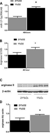
miR-17-5p and arginase II levels are increased by hypoxia. A: hypoxia increased miR-17-5p expression in human pulmonary artery smooth muscle cells (hPASMC). hPASMC were exposed to normoxia or hypoxia for 48 h (n = 3 for each group). miR-17-5p levels were analyzed by quantitative real-time PCR and normalized to RNU48 expression. Data are shown as means ± SE relative to respective normoxia controls at each time point. *Hypoxia different from normoxia controls, P < 0.05. B: hypoxia induces the increase in arginase II mRNA expression in hPASMC. hPASMC were exposed to normoxia or hypoxia for 48 h (n = 3 for each group). Arginase II mRNA levels were analyzed by quantitative real-time PCR and normalized to 18S expression using the ΔΔCT method. Data are shown as means ± SE relative to respective normoxia controls. *Hypoxia different from normoxia controls at same time point, P < 0.05. C: hypoxia induces an increase in arginase II protein expression in hPASMC. hPASMC were exposed to normoxia or hypoxia for 48 h (n = 3 for each group). Representative Western blots are shown for arginase II and β-actin. D: densitometric analysis of arginase II protein expression levels normalized to β-actin. Data are shown as means ± SE relative to β-actin. *Hypoxia different from normoxia controls at same time point, P < 0.05.