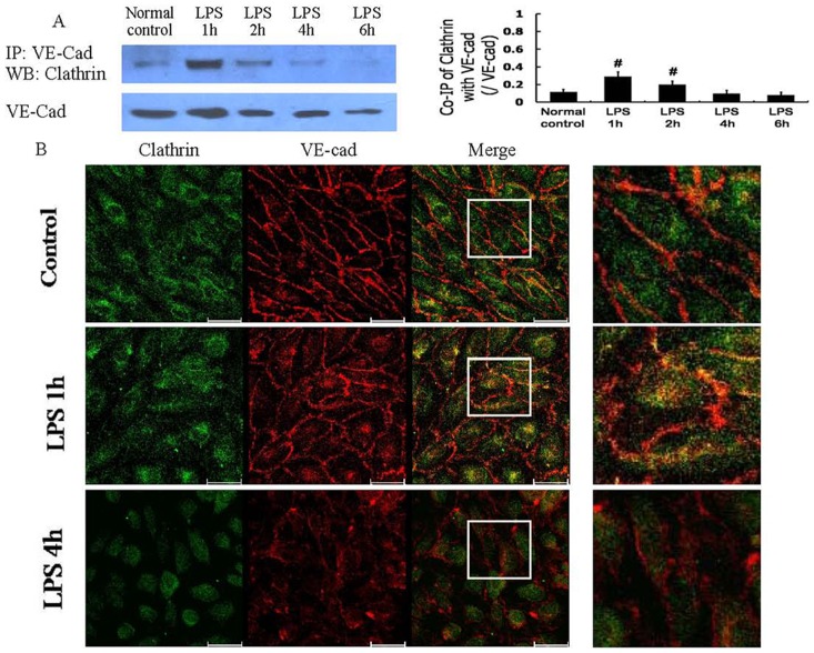
Figure 2. Clathrin-mediated endocytosis of VE-cad after LPS treatment.
The co-immunoprecipitation of VE-cad with clathrin was dominant 1 h after LPS treatment (A); immunocytochemistry and confocal microscopy observations also demonstrated the significant co-localization (yellow) of VE-cad with clathrin 1 h after LPS treatment (B). Scale bars: 40 µm. # P<0.05 vs normal control group.

