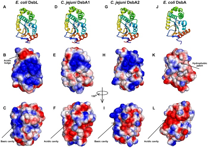
Figure 2. Homology models of C. jejuni DsbA1 and DsbA2.
C. jejuni DsbA1 and DsbA2 (CjDsbA1 and CjDsbA2) models built on E. coli DsbA [EcDsbA (PDB ID: 2ZUP [80])] and DsbL [EcDsbL (PDB ID: 3C7M [22])], experimentally characterized members of the DsbA superfamily. Structural representations are shown in ribbon view (A, D, G and J). Electrostatic surfaces coloured by charge from red, acidic, -1kT to blue, basic, +1kT. The orientation in B, E, H and K follows the orientation in the top row (A, D, G and J) and in C, F, I and L is rotated by 130 degrees around the vertical axis, clockwise.

