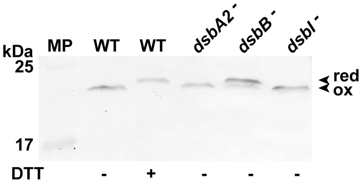
Figure 8. Redox state of CjDsbA1 in C. jejuni 81116 strains: wild type (WT), cjdsbA2-, cjdsbB- and cjdsbI- mutants.
Bacterial cultures were treated with 10% TCA, followed by alkylation with AMS (4-acetamido-4′-maleimidylstilbene-2,2′-disulfonic acid). Cellular proteins including the reduced (red; DTT treated, modified by AMS) controls were separated by 15% SDS-PAGE under non-reducing conditions, followed by Western blot analysis using antibodies against CjDsbA1. Each lane contains proteins isolated from the same amount of bacteria. The relative positions of protein molecular weight standard (MP) are listed on the left (in kilodaltons). The figure presents a representative result.

