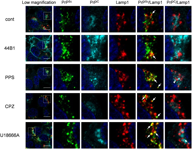
Figure 7. Co-localization of PrPSc or PrPC with Lamp1.
ScN2a-3-22L cells were cultured under the same condition as described in Figure 6. The cells were subjected to direct immunostaining of PrPC and PrPSc with 31C6-Af555 and rIgG132-EGFP, respectively and subsequently to immunostaining for Lamp1 and nuclei. The leftmost column shows a lower-magnification merged image of PrPSc (green), PrPC (cyan), Lamp1 (red), and nuclei (blue). Individual and merged high-magnification images of the boxed regions are shown on the right. Arrows or arrowheads denote representative examples of the co-localization of PrPSc with Lamp1 or PrPC with Lamp1, respectively. Scale bars: 10 µm.

