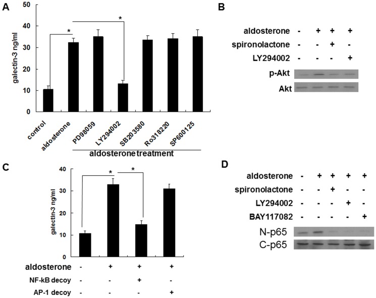
Figure 5. Aldosterone induced galectin-3 expression in THP-1 cells via a MR/PI3K/Akt/NF-κB signaling pathway.
(A) Aldosterone induced galectin-3 expression in THP-1 cells via the PI3K/Akt signaling pathway. THP-1 cells were serum-starved for 24 h and then pre-treated with PD98059 (50 µg/ml), LY294002 (50 µg/ml), SB203580 (5 µg/ml), Ro318220 (5 nM), and SP600125 (10 µM) for 1 h prior to 10−6 M aldosterone or vehicle treatment for 24 h. (B) Aldosterone induced Akt phosphorylation via MR-mediated signaling. THP-1 cells were serum-starved for 24 h, then pre-treated with different chemical inhibitors for 1 h prior to 10−6 M aldosterone or vehicle treatment. After 30 minutes, total protein was collected and the expression level of the indicated protein was measured by Western blot with specific antibodies. (C) NF-κB was a prerequisite transcriptional factor in aldosterone-mediated galectin-3 expression. THP-1 cells were transfected with NF-κB decoy ODN, AP-1 decoy ODN and serum starved for 24 h, then treated with 10−6 M aldosterone for another 24 h. * P<0.05. (D) Aldosterone activated NF-κB via MR/PI3K/Akt signaling. THP-1 cells were serum-starved for 24 h and then pre-treated with 10−7 M of spironolactone, LY294002 (50 µg/ml) or BAY117082 (10 µM) for 1 h before 10−6 M aldosterone treatment. Nuclear and cytosolic proteins were collected after 1 h, and the level of the NF-κB p65 subunit was determined by Western blotting. MR = mineralocorticoid receptor. nuclear (N) p65 and cytosol (C) p65.

