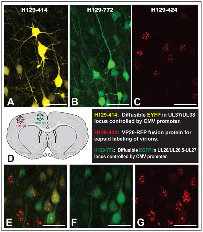
Figure 10.

Efficiency of reporter gene expression and the utility of H129 recombinants for dual tracing studies are illustrated. The 414 and 772 reporters generate diffuse cytoplasmic fluorophores that densely stain the somatodendritic compartment of infected neurons (A & B). The 424 recombinant expresses mRFP as part of a fusion protein with the UL35 (VP26) gene under the control of its native promoter. VP26 is a capsid protein so mRFP appears as punctate labeling within infected neurons (C). Furthermore, the distribution of punctate mRFP labeling is reflective of the stage of viral replication such that early labeling is characterized by differential concentration of mRFP in cell nuclei and the subsequent appearance of mRFP in the cell soma, dendrites and axons occurs with advancing viral replication. Figure D illustrates one of three dual infection paradigms included in the analysis, each of which involved recombinant 424 in combination with either 414 or 772. In each paradigm anterograde transneuronal spread of recombinants resulted in the dual infection of neurons that were easily identified due to the differential concentration of reporters in the nucleus versus the cell cytoplasm (E – F). Marker bars A – G = 50 μm.
