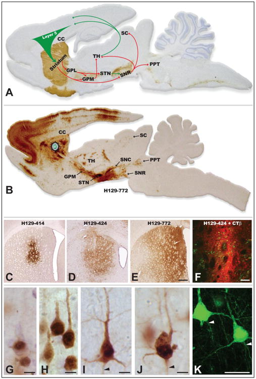
Figure 5.

The organization of basal ganglia circuitry used to characterize the invasiveness of H129 recombinants is illustrated in A. Tyrosine hydroxylase was localized in the sagittal section using immunoperoxidase procedures (brown reaction product) to highlight the nigrostriatal dopaminergic projection that is a major component of the circuit and the section was counterstained with cresyl violet. Arrows indicate the direction of projection pathways that would produce anterograde transneuronal passage of virus after injection of virus into striatum. Green arrows indicate excitatory projections and red arrows inhibitory projections. Figure B illustrates the distribution of viral infection 72 hours after injection of H129 recombinant 772 into the striatum. The sagittal section shows a plane of section similar to that illustrated in A and infected cells were localized using an anti-HSV antiserum and immunoperoxidase procedures. Images C – F illustrate the distribution of infected neurons in the striatum 72 hours after injection of H129 recombinants into the striatum, either alone (C – E) or in combination with the classical tracer CTβ. Note the variability of the intrastriatal spread of infection produced by recombinants 414, 424 and 772. The primary site of injection is marked by the dense deposit of CTβ (red fluorescence), which distinguishes the zone of first order infection from transneuronal spread of virus (green fluorescence localizes infected cells). Stages of viral infection illustrative of the temporal kinetics of viral infection are illustrated in images G – K. Image K illustrates viral antigen localized with immunofluorescence and imaged with confocal microscopy. Infected cortical pyramidal neurons early in viral replication demonstrate differential concentration of viral antigens within the cell nucleus (G). At intermediate stages of infection (H) the dense staining of the nuclei of infected pyramidal neurons is complemented by the appearance of viral antigens in cytoplasm of cell somas and primary dendrites. With advancing replication (I – K) viral antigen fills the entire somatodendritic compartment and is also evident within the initial segments of axons (arrowheads). Cytopathology is also evident in the longest infected neurons, and is characterized by swelling of the cell body, displacement and invagination of the cell nucleus within the cell soma, and occasional beading of dendrites (J). Abbreviations: cc = corpus callosum, GPL = lateral subdivision of globus pallidus, GPM = medial subdivision of globus pallidus, PPT = pedunculopontine tegmental nucleus, SC = superior colliculus, SNC = substantia nigra, pars compacta, SNR = substantia nigra, pars reticulata, STN = subthalamic nucleus, TH = thalamus. Marker bar in E = 500 μm and images C – E are of the same magnification, marker bar in F = 100 μm, marker bars in G – J = 10 μm, marker bar in K = 25 μm.
