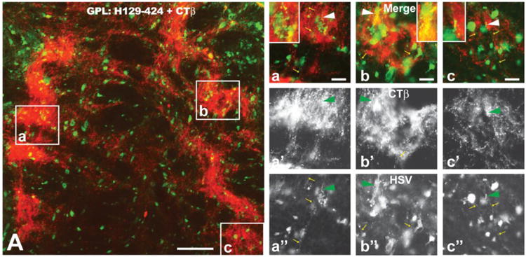
Figure 6.

The distribution of viral antigens (green fluorescence) in relation to CTβ - labeled (red fluorescence) striatal axons in the lateral subdivision of the globus pallidus (GPL) is illustrated 72 hours after injection of a cocktail of CTβ and H129 recombinant 424 into the dorsomedial quadrant of the striatum. The GPL is shown in sagittal section with the left border of image A representing the rostral pole of GPL. Note the dense termination of striatal afferents within subregions of GPL. Infected neurons coextensive with striatal afferents were likely labeled by first-order anterograde transneuronal spread of virus. Infected neurons outside of fields containing labeled striatal afferents were likely labeled by anterograde spread of the recombinant within striatum and subsequent anterograde spread to GPL. The boxed areas in image A are shown at higher magnification in a, b and c. Images a′, b′ & c′ and a″, b″ & c″ show only the red (CTβ) or green (viral antigen) channels in a, b & c. Note that CTβ labeled afferents rarely contain viral antigens (arrowhead a – c″ and insets of a - c) but that axons of local circuit neurons or recurrent collateral of infected GPL neurons (small yellow arrows in a – c and a″ – c″) are immunopositive for viral antigens. See text for a more detailed explanation. Marker bar in figure A = 100 μm; the marker bars in a, b and c = 20 μm and the corresponding images of the red and green channels are of the same magnification.
