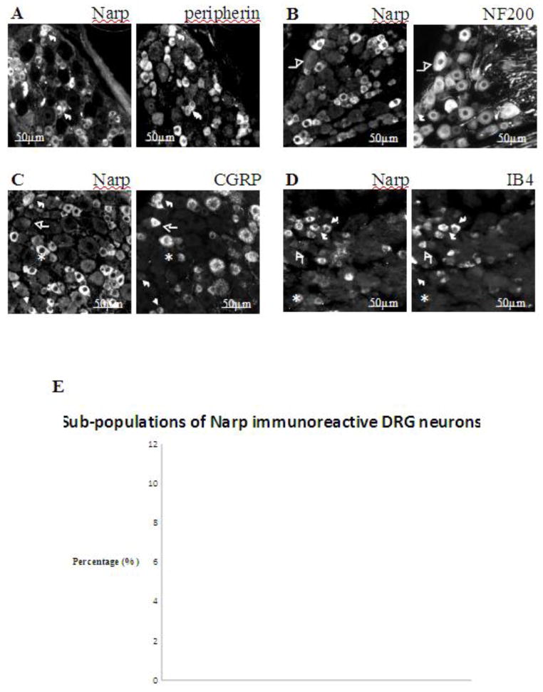
Figure 2.
Characterization of Narp immunoreactive DRG neurons. Fluorescent photomicrographs of naïve mouse lumbar (L4 to L6) DRG show Narp double-staining with peripherin (A), NF200 (B), CGRP (C), and IB4-binding (D). Short arrows highlight neurons that co-express Narp and marker. Asterisks highlight neurons that only express Narp. Long arrows highlight neurons that only express marker. (E) Mean percentages of Narp immunoreactive DRG neurons expressing each of these markers.

