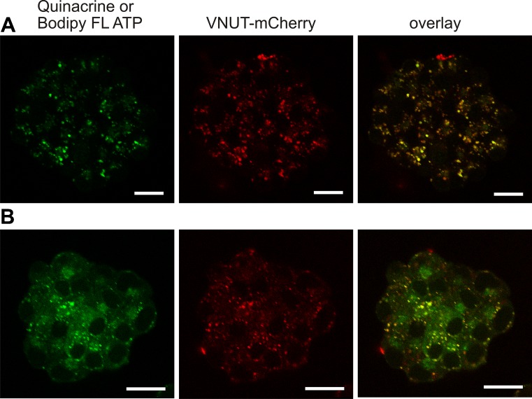
Fig. 2.
Co-localization of VNUT-mCherry and markers of ATP stores in living cells. AR42J cells were transfected with VNUT-mCherry, and cells were incubated either with 5 μM quinacrine (first row) (a) or 15 μM Bodipy FL ATP (second row) (b). The following excitation/emission wavelengths were used: VNUT-mCherry 594/600–700 nm (red), quinacrine 458/466–578 nm (green), and Bodipy FL ATP 488/500–550 nm (green); respective red and green images and overlay images are shown. Representative images are of three independent experiments. Scale bars are 25 μm

