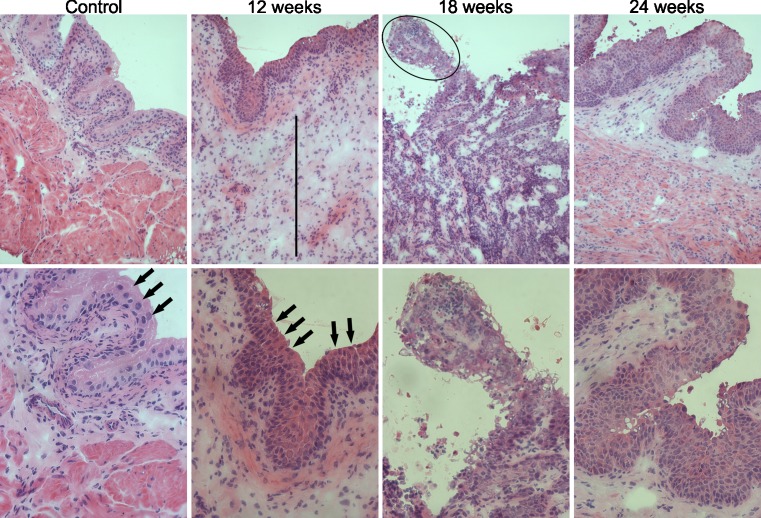
Fig. 2.
The images correspond to hematoxylin and eosin staining of bladders from control animal and bladders from animals which received BBN for 12, 18, and 24 weeks, correspondent to each column respectively. Images of the top row were taken with ×200 of increase and the images of the second row with ×400. Black arrows indicate examples of umbrella cells, specific from urothelium. It could be observed that the bladder from animals that received BBN for 12 weeks present features of inflammation, mainly edema (black line indicates the length of edema area). Cancer features as a papillary carcinoma (indicated by black circle) and loss of umbrella cells, increase of number of cells and cell crowding, could be observed in bladders from 18 to 24 weeks

