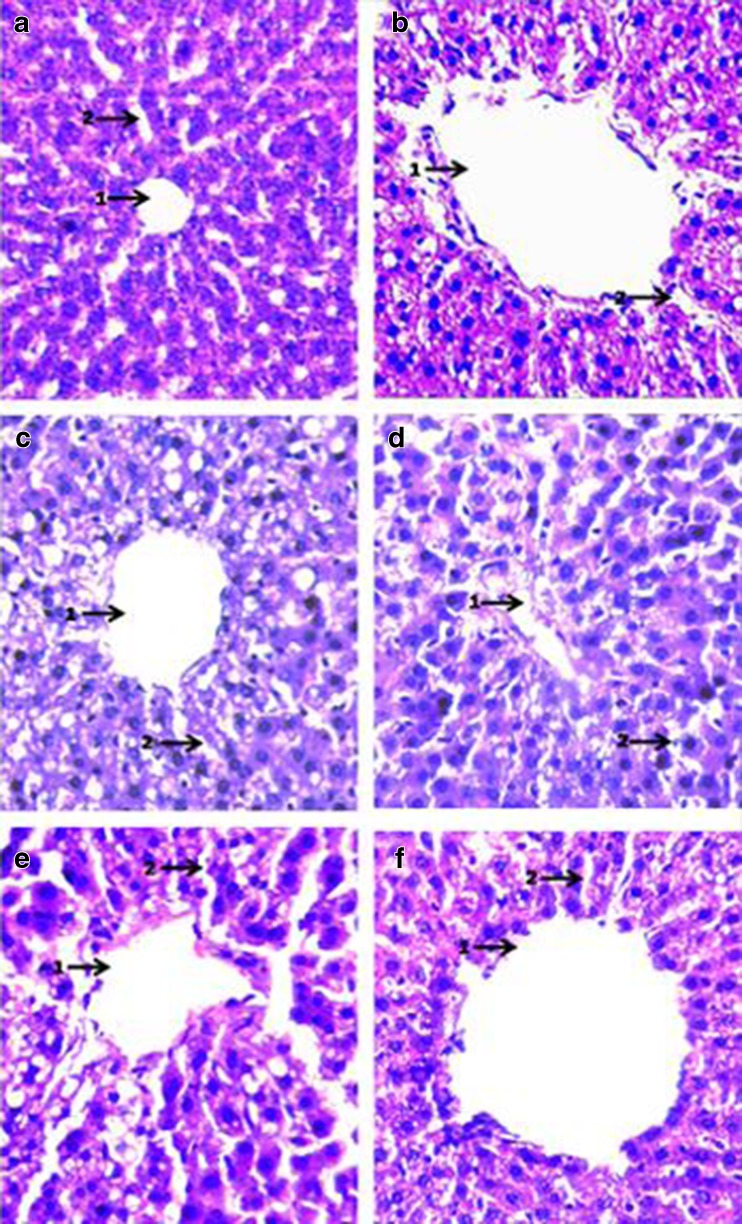
Fig. 7.
Histopathological examination of Haematoxylin-Eosin stained sections of liver (magnification × 400): a Negative control (SO fed + non-diabetic): 1. Patent central vein surrounded by well defined hepatocytes; 2. Sinusoids well defined. b Positive control (SO fed in alloxan induced diabetic rats) 1. Leaky central vein (extremely distended) surrounded by ruptured hepatocytes; 2. Distorted sinusoids. c SO + (0.5 % ESA) NE fed in alloxan induced diabetic rats: 1. Central vein significantly reclaimed with slightly enlarged hepatocytes; 2. Sinusoids well defined in major areas. d SO + (0.25 % ESA) NE fed in alloxan induced diabetic rats: 1. Central vein comparable with normal control surrounded by regular hepatocytes; 2. Sinusoids well defined. e SO + (0.5 % ESA) CE fed in alloxan induced diabetic rats: 1. Enlarged hepatocytes surrounding slightly distorted central veins; 2. Ruptured sinusoids. f SO + (0.25 % ESA) CE fed in alloxan induced diabetic rats: 1. Distended central vein surrounded by enlarged hepatocytes; 2. Sinusoids non-regular

