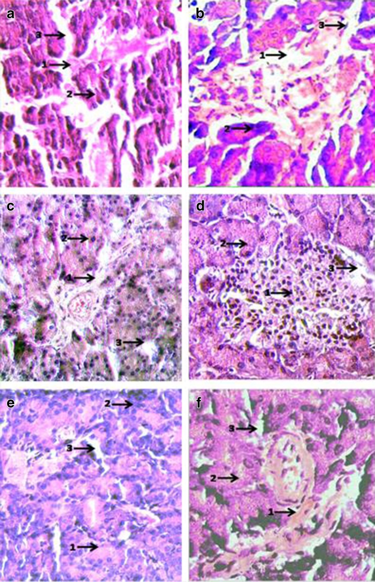
Fig. 8.
Histopathological examination of Haematoxylin-Eosin stained sections of Pancreas (Magnification × 400): a Negative control (SO fed + non-diabetic): 1. Patent Islets of Langerhans (weakly stained); 2. Normal acinar cells (dark stained); 3. Regular blood Capillaries and vessels. b Positive control (SO fed in alloxan induced diabetic rats): 1. Ruptured Islets of Langerhans; 2. Ruptured and enlarged acinar cells; 3. Distended and ruptured blood vessels. c SO + (0.5 % ESA) NE fed in alloxan induced diabetic rats: 1. Reclaimed Islets of Langerhans; 2. Acinar cells with enlarged nuclei; 3. Open and patent blood vessels. d SO + (0.25 % ESA) NE fed in alloxan induced diabetic rats: 1. Completely reclaimed Islets of Langerhans with leukocyte infiltration in patches; 2. Regular/normal acinar cells; 3. Open and patent blood vessels. e SO + (0.5 % ESA) CE fed in alloxan induced diabetic rats: 1. Islets of Langerhans restituted; 2. Enlarged acinar cells; 3. Normal blood vessels. f SO + (0.25 % ESA) CE fed in alloxan induced diabetic rats: 1. Islets of Langerhans not restored completely; 2. Acinar cells not well defined; 3. Distorted blood vessels

