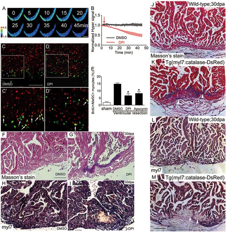
Figure 2.
H2O2 signaling is required for heart regeneration. (A-B) Inhibiting Duox/Nox NADPH oxidases by DPI decreased H2O2 generation. Time-lapse confocal images (A) and statistics (B) of the Hyper ratio in Tg(myl7:Hyper) hearts prior to and after application of DPI (10 μM). DMSO (0.1%) was used as control. n = 3-5. (C-E) Treatment with DPI or apocynin impaired cardiac myocyte regeneration. Proliferating myocytes were identified by double-staining with anti-Mef2C (red) and anti-BrdU (green) (C', D', arrows). Note that there were fewer double-positive cells (yellow) in DPI-treated heart (D, D') than in DMSO control heart (C, C'). Quantitative results with sham, DMSO, DPI or apocynin treatment are shown in E. n = 5 to 7. See Materials and Methods for details of treatment. (F-I) Accumulated fibrin/collagens (white arrowheads in G, Masson's staining) and compromised myocardial regeneration (white arrowhead in I, in situ hybridization with myl7 probes) after DPI treatment, compared with DMSO control (F, H). (J-M) Cardiac-specific overexpression of catalase retarded heart regeneration. Tg(myl7:Catalase-DsRed) heart displayed larger amounts of fibrin/collagens (white arrowheads in K, Masson's staining) and compromised myocardial regeneration (white arrowheads in M, in situ hybridization with myl7 probes), as compared with respective non-transgenic sibling hearts at 30 dpa (J, L). n = 5-8. Scale bars, 100 μm.

