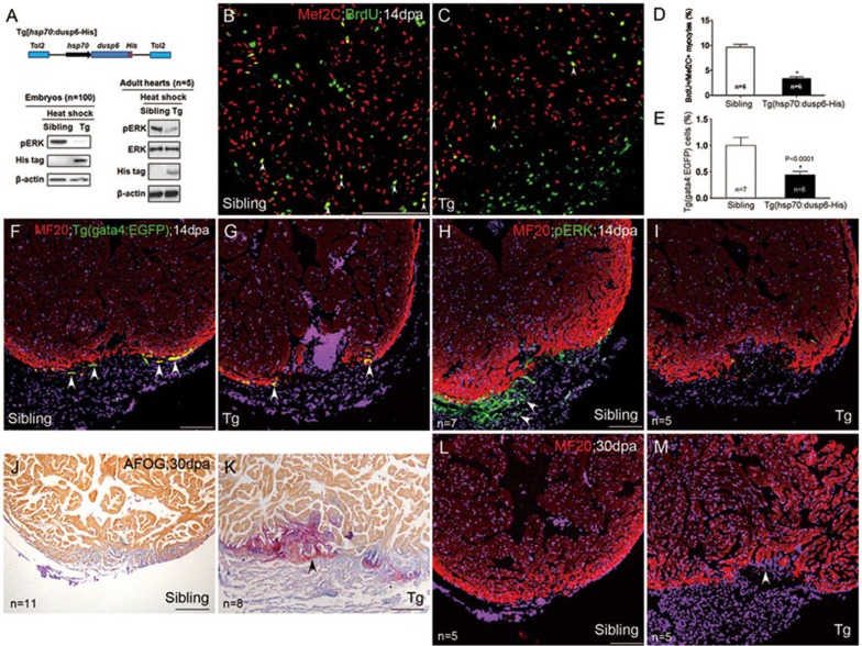
Figure 7.
The repressive effects of Dusp6 overexpression on heart regeneration through dephosphorylation of pERK. (A) Top: a scheme showing that in Tg(hsp70:dusp6-His), His-tagged Dusp6 is driven by the zebrafish hsp70 promoter. Transgenesis was mediated by Tol2 transponase. Bottom: heat shock-induced expression of His-tagged Dusp6 proteins diminished pErk in transgenic embryos (Tg) at 72 hpf or transgenic adult hearts (Tg), compared with their sibling embryos or adult hearts. β-actin was used to normalize protein loading. (B-D) Mef2C+/BrdU+ proliferating myocytes (arrowheads) were decreased in dusp6 transgenic hearts (C) compared with those in non-transgenic siblings (B), with the statistics shown in D. (E-G) Representative images showing that Tg(gata4:EGFP) myocytes (arrowheads), which colocalized with cardiac marker MF20, were diminished in dusp6 transgenic hearts (G) compared with their siblings (F), with the statistics shown in E. (H-I) Reduction of pErk+ cells (arrowheads), not overlapping with cardiac marker MF20, in transgenic hearts (I), compared with non-transgenic siblings (H). (J-M) Transgenic overexpression of Dusp6 led to cardiac fibrosis (assayed by AFOG staining, K) and compromised cardiac regeneration (assayed by MF20 staining, M), compared with their siblings (J, L). Nuclei were co-stained with DAPI. Scale bars, 100 μm.

