Figure 2.
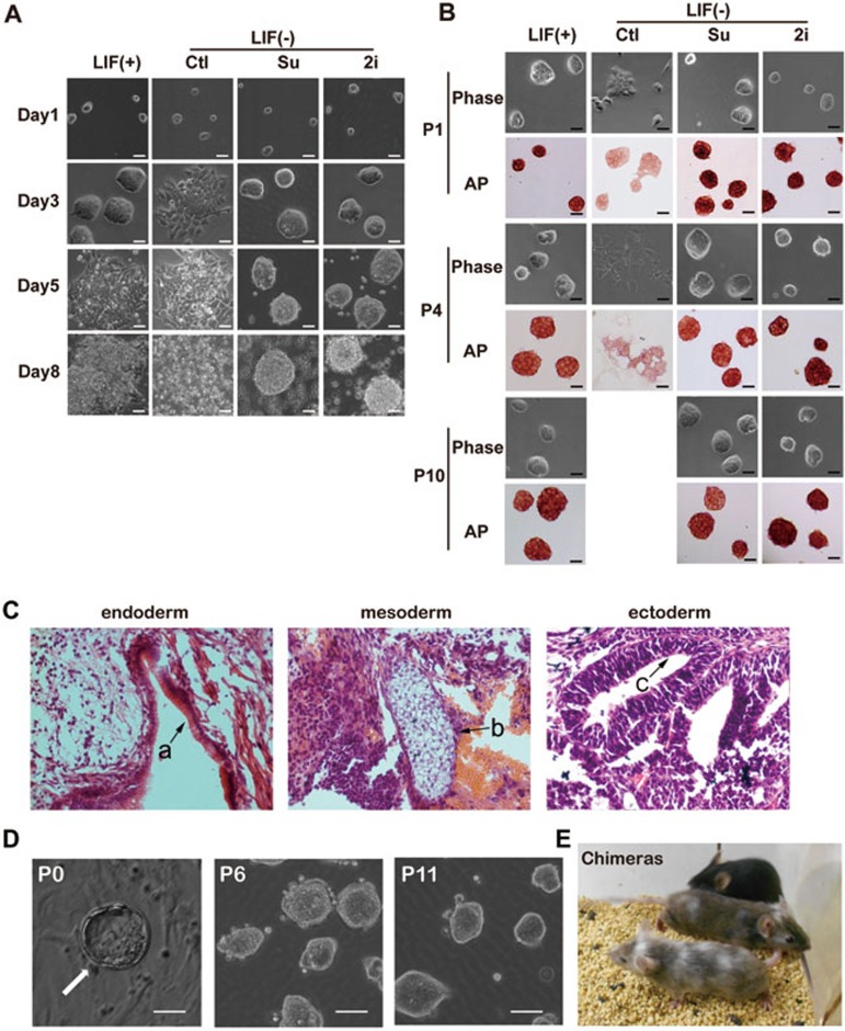
Sunitinib maintains the self-renewal and pluripotency of mESCs. (A) Morphology of E14 cells cultured in mES media containing 1 000 U/ml LIF, or in mES media without LIF but containing 2i (3 μM PD0325901 and 3 μM CHIR99021) or sunitinib (Su, 1 μM), for 1-8 days. Media were changed every day. Scale bar, 50 μm. (B) Morphology and alkaline phosphatase (AP) staining of E14 cells at passages 1, 4 and 10 maintained in mES media containing 1 000 U/ml LIF, or in mES media without LIF but containing 2i (3 μM PD0325901 and 3 μM CHIR99021) or sunitinib (1 μM). Scale bar, 50 μm. (C) H&E staining of teratomas generated with E14 cells maintained with sunitinib (passage 11). Typical structures of the three embryonic germ layers are shown as follows: a, gastrointestinal-like epithelium (endoderm); b, cartilage-like structure (mesoderm); c, neural tube-like structure (ectoderm). Scale bars, 20 μm. (D) Blastocyst outgrowth (P0, the arrow) on feeder cells and mES medium containing sunitinib and 2 000 U/ml LIF. mESCs were subsequently maintained in mES media containing sunitinib for multiple passages. Scale bar, 20 μm. (E) Chimeric mice generated with mESCs derived and maintained with sunitinib (passage 6).

