Figure 1.
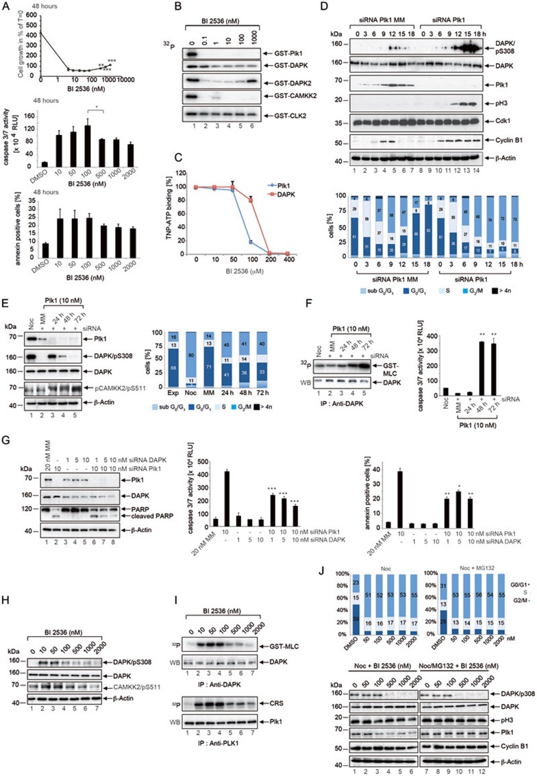
Characterization of the mitotic roles of DAPK in Plk1-inihibited HeLa cells. (A) Concentration-dependent proliferative and apoptotic responses upon BI2536 treatment. (B) Evaluation of novel BI2536 targets using in vitro kinase assays. The purified proteins were subjected to kinase assays and the autophosphorylation activities were monitored using γ-32P ATP. (C) TNP-ATP displacement assay for the analysis of BI2536 binding to Plk1 and DAPK. (D) Cells treated with Plk1 siRNA or Plk1-MM siRNA were arrested in G1/S by double thymidine-treatment. At the indicated time points, cells were lysed and immunoblotted for DAPK/pS308, DAPK, Plk1, pH3, Cdk1, Cyclin B1 and β-Actin (upper panel). Cell cycle was analyzed by FACS at the indicated time points (lower panel). (E) Cells were treated with nocodazole for 16 h or transfected with Plk1-MM siRNA as control for 24 h or Plk1 siRNA for 24 h, 48 h and 72 h. Lysates were immunoblotted for Plk1, DAPK/pS308, DAPK, CAMKK2/pS511 and β-Actin (left panel). Cells were analyzed by FACS at the indicated time points (right panel). (F) DAPK immunoprecipitated from the lysates of siRNA-treated cells was subjected to kinase assays using GST-MLC as the substrate (left panel) and caspase-3/7 activity was determined (Caspase-Glo 3/7 Assay) (right panel). **P < 0.01, Student's t-test, unpaired and two-tailed. (G) HeLa cells were transfected with siRNA as indicated. Twenty-four hours after transfection, cell lysates were analyzed by immunoblotting for Plk1, DAPK, PARP and β-Actin (left panel). Caspase-3/7 activity was determined (middle panel) and 7-AAD was used in conjunction with annexin V staining to discriminate among the viable, apoptotic and necrotic cells using dual parameter FACS analysis (right panel). *P < 0.05, **P < 0.01, ***P < 0.001, Student's t-test, unpaired and two-tailed. (H) Lysates of cells treated with increasing concentrations of BI2536 for 24 h were immunoblotted for DAPK/pS308, DAPK, CAMKK2/pS511 and β-Actin. (I) DAPK and Plk1 immunoprecipitated from lysates were subjected to kinase assays using GST-MLC and cytoplasmic retention signal (CRS) of cyclin B1 as the substrates, respectively. (J) Cells enriched in mitosis by nocodazole- or nocodazole/MG132-treatment were incubated with increasing concentrations of BI2536 for 24 h. Cell cycle analyses by FACS (upper panel) and immunoblotting (lower panel) were then performed.

