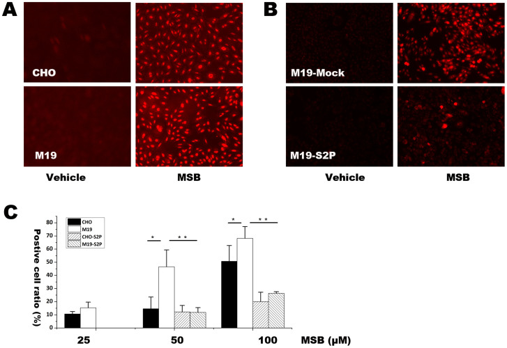
Figure 3. HEt staining followed by fluorescence microscopic observation and flow cytometery for determining superoxide level after treatment with MSB in CHO, M19, M19 cells transfected with Mock and S2P vector.
(A) Fluorescent picture of HEt staining in CHO and M19 cells treated with 20 μM MSB for 2 h. (B) Fluorescent picture of HEt staining under treatment of 20 μM MSB for 2 h in M19 cells transfected with Mock or S2P vector. (C) Percentages of HEt positive cells were determined by flow cytometery in CHO and M19 cells with or without transfection of S2P gene. The percentages of positive staining cells were expressed as mean ± SD, n = 3. Statistical analysis was done by ANOVA. * p < 0.05, ** p < 0.01. The results showed that S2P reduced superoxide production induced by MSB.

