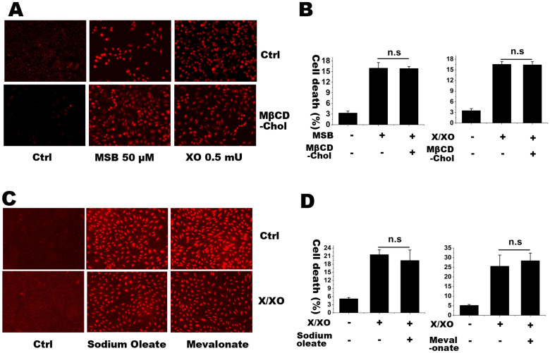
Figure 5. HEt staining and LDH release assay for detecting superoxide level and the rates of cell death, respectively, in M19 cells with or without water soluble cholesterol MβCD-chol, sodium oleate and mevalonate.
(A) is the superoxide detection in M19 cells treated with MSB (50 μM) or X/XO (X, 100 μM; XO, 0.5 mU) with or without 50 μM MβCD-chol for 2 h. (B) is the rate of cell death with the same treatments. (C) is superoxide detection in M19 cells treated with X/XO (X, 100 μM; XO, 0.5 mU) with or without 1 mM sodium oleate and 100 μM mevalonate for 2 h. (D) is the rate of cell death with the same treatments. The percentages of cell death were expressed as mean ± S.D. n = 3. Statistical analysis was done by ANOVA. n.s.: no significance. The results showed that lipids incorporation did not affect cellular superoxide level and cell death induced by MSB and X/XO.

