Figure 7.
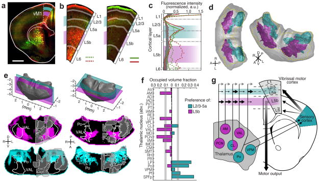
Cortical layer preferences of thalamic projections to primary vibrissal motor cortex (vM1). (a) Coronal brain section showing layers 2/3 and 5a (L2/3–5a, cyan) and layer 5b (L5b, magenta) in vM1 (white dashed outline). (b) Example coronal sections of vM1 (left image magnified from panel a), showing thalamocortical projections with preference to L5b (red projections, left), and L2/3–5a (red and green projections, right). (c) Normalized fluorescence intensity plots for red and green projections in panel b (left image: dashed lines, right image: solid lines). Fluorescence is averaged radially across vM1 to determine layer preference (see Methods and Supplementary Fig. 14). Background fluorescence is calculated from brains without vM1 projections (gray line). (d) Dorsal (left) and oblique (right) views of a 3D thalamus rendering, showing the volumes that are associated with preferential axonal projections to L2/3–5a (cyan) and L5b (magenta). The total volume projecting to all layers of vM1 is shown (gold dashed line). (e) Representative coronal (left) and horizontal (right) sections of modified confidence maps for L2/3–5a (cyan) and L5b (magenta) preferential vM1 projections. Outlines of thalamic nuclei are overlaid on each section image. (f) The occupied fraction of each thalamic nucleus containing layer-preferential projections to L2/3–5a and 5b of vM1. An arbitrary 10% threshold is indicated (gray line). (g) Schematic showing layer preferential input from thalamus to vM1 in the context of a motor-sensory circuit diagram. All scale bars are 1 mm.

