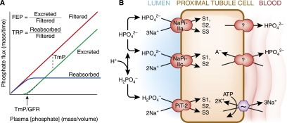
Figure 4.

Proximal tubule phosphate transport. (A) Concepts of renal inorganic phosphate (Pi) homeostasis by the proximal tubule. The flux of filtered and reabsorbed Pi is plotted against plasma phosphate concentration; the difference between the two yields the rate of excretion of Pi. There are a number of terms used to quantify the proximal tubule’s Pi reabsorption at the whole organism level. Fractional excretion of Pi (FEP) and tubular reabsorption of Pi (TRP) sum to unity (FEP=1−TRP). The maximal tubular reabsorptive capacity of Pi (TmP in units of mass/time) refers to the saturating transepithelial flux of Pi that the tubule can mount and is equal to the difference between filtered and absorbed phosphate when the filtered load is higher than TmP. The plasma concentration threshold at which Pi starts to appear in the urine is TmP/GFR (in units of mass/volume). (B) Cell model of proximal tubule Pi transport. Three apical transporters mediate Pi entry with different preferred valence of Pi, stoichiometry of Na+, electrogenicity, and pH gating. The affinities for Na+ are all approximately 30–50 mM but are much higher for phosphate (0.1, 0.07, and 0.025 mM for NaPi-Ila, NaPi-Ilc, and PiT-2, respectively). Distribution in the proximal tubule segments (S1, S2, S3). Basolateral Pi exit occurs via unknown mechanisms. Apical Na-coupled Pi transport is inhibited in acidosis by alteration in luminal substrate, directly gating of the transporter by pH, and decreased apical NaPi transporters as described in Figure 3B.
