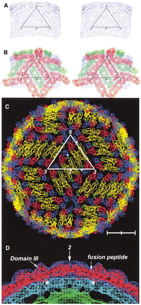
Figure 3. Fit of E Dimers into Density.

(A) and (B) correspond to density that is between planes, perpendicular to an icosahedral 2-fold axis and at a distance of 220-250 A from the viral center. The contour level is at about 4σ (root mean square deviation from the mean density).
(A) Stereo diagram showing only the density between 220 and 245 A radius.
(B) Stereo diagram showing the interpreted density in terms of dimers on icosahedral 2-fold axes (green) and dimers on quasi-2-fold axes (red).
(C) Structure of the whole virus showing each monomer with domains I, II, and III in red, yellow, and blue, respectively. The fusion peptide is shown in green. The C-terminal residue 395 is shown as a white asterisk for monomers within the defined icosahedral asymmetric unit. Note the pair of holes in each dimer. Scale bar represents 100 A.
(D) Central crosssection through the cryo-EM density showing the outer radial shells as in Figure 1B. The direction of the crosssection follows the length of a dimer situated on an icosahedral 2-fold axis. The arrow indicates the position of the 2-fold axis. The location of domain III and the fusion peptide in domain II are shown. Asterisks indicate the carboxy end of the fitted E protein.
