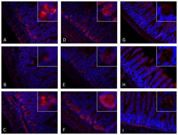
Figure 1. Representative immunohistochemistry (IHC) of antimicrobial peptides in ileum from Chow, PN, and PN+BBS-fed mice.
IHC of sPLA2 in A) Chow-fed mice; B) PN-fed mice; C) PN+BBS-fed mice. IHC of lysozyme in D) Chow-fed mice; E) PN-fed mice; F) PN+BBS-fed mice. IHC of RegIII-γ in G) Chow-fed mice; H) PN-fed mice; I) PN+BBS-fed mice. Images are 20X magnification of intestinal villi and crypts. Staining for sPLA2 and lysozyme is decreased in PN-fed mice and appears qualitatively similar in Chow and PN+BBS-fed mice. RegIII-γ staining appears cytosolic and localizes around intestinal villi. PN, parenteral nutrition; BBS, bombesin.

