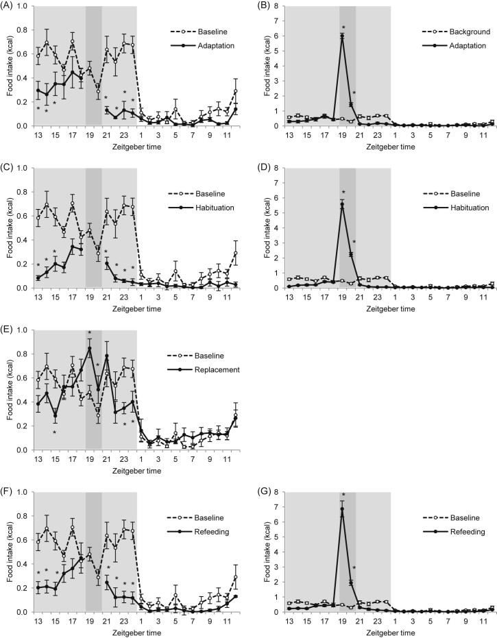
Fig. 2.
Food intake microstructure of C57BL/6 mice during all study phases versus baseline food intake pattern: (A,B) adaptation, (C,D) habituation, (E) replacement and (F,G) refeeding phase. (A,C,E,F) Food intake microstructure showing caloric intake from standard diet. (B,D,G) Food intake microstructure showing total caloric intake including calories from HFD during scheduled feeding. Light shaded area indicates dark phase; dark shaded area indicates scheduled feeding time. *P < 0.05 versus baseline by two-way repeated measures ANOVA and Student–Newman–Keul post hoc test. For clarity, one asterisk also includes P < 0.01 and P < 0.001, and diagrams (B,D,G) display only differences during scheduled feeding time versus baseline (ZT19 and ZT20). Data are presented as mean ± SEM.

