Fig. 3.
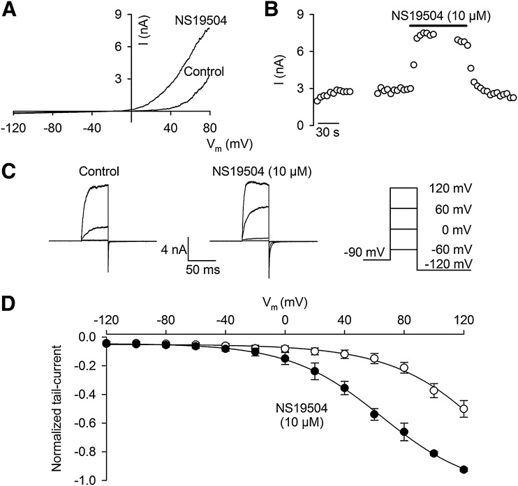
Electrophysiological characterization and activation of BK channels by NS19504. Current was measured in inside-out patches obtained from HEK293 cells stably expressing BK channels. Experiments were conducted at a physiologic K+ gradient (4/154 mM K+) and at a free intracellular Ca2+ concentration of 0.3 µM. (A) Current-voltage (I-V) relationships measured in the absence (Control) or presence of 10 µM NS19504 in the intracellular/bath solution. Currents were elicited by applying linear voltage ramps from −120 to +80 mV from a holding potential of −90 mV. (B) BK current measured at +80 mV depicted as a function of time. NS19504 (10 µM) was applied to the inside of the patch, indicated by the bar. Breaks in the recording indicate periods where voltage step protocols were applied. Data are from a single experiment representative of four independent experiments. (C) BK current activated by membrane potential steps, as illustrated in the drawing to the right. (D) Normalized tail currents depicted as a function of step potential. Data are mean ± S.E.M. of four independent experiments. Peak tail currents were measured by stepping to −120 mV after obtaining steady current activation at depolarized potentials either in the absence or presence of 10 µM NS19504.

