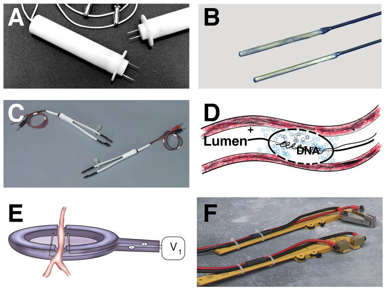
Figure 2. Electroporator probes and electrodes.
A number of different electrodes for in vivo electroporation-mediated gene transfer have been developed. Some electrodes can be used for multiple tissue applications (A, B, C, and F), whereas others are limited to certain tissues, such as the vasculature (D and E). Two-needle electrodes (A), “Genetrode” rod electrodes (B), “Tweezertrode” tweezer electrodes (C), porous balloon-catheter electrode (25, 26)(D), spoon electrode (79)(E), and caliper electrodes (F) are shown. Electrodes shown in A, B, C, and F are from BTX (Harvard Instruments). The small blue stars in panel D represent plasmids.

