
Figure 4. Sequence-specific nuclear localization and GFP expression.
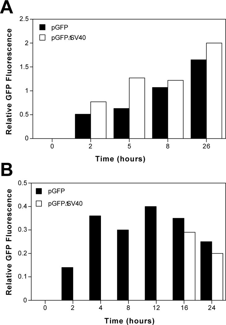
(A) Nuclear injected pGFP and pGFPΔSV40 display similar patterns and efficiencies of GFP expression. TC7 cells were synchronized by treatment with 60 μM lovastatin for 16 hours, washed and then incubated in medium containing serum and 5 mM mevalonic acid for two hours before microinjection. Thirty copies of either plasmid were injected into each nucleus of approximately 100–150 cells and assayed for GFP expression at later times. GFP expression which was recorded on an intensity scale of 1 to 5 and relative GFP expression is given as the total intensity divided by the total number of injected cells. (B) Plasmids with or without the SV40 enhancer differ greatly in their ability to express GFP when injected into the cytoplasm. Thirty copies of either pGFP or pGFPΔSV40 were microinjected into the cytoplasm of approximately 100 cells, synchronized as in (A), and relative GFP expression was measured. The results shown (A and B) are representative of 4 independent experiments.